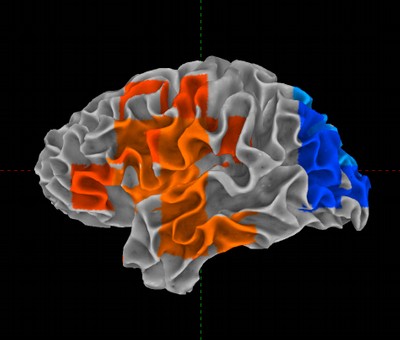
Rendered brain showing abnormal brain activity in torture victims.
This picture (white-gray matter border) shows regions in red with excess slow wave activity which is strongest in the left insula (see Kolassa et al. "Imaging the trauma: altered cortical dynamics after repeated traumatic stress") and the left frontal inferior region (see Ray, William and Thomas Elbert "Survivors of organized violence often left with traumatic memories." Psychological Science Volume 17, Issue 10, October 2006). Blue indicates less activity than normal (N = 97 / group). Photo: Courtesy of Dr. Thomas Elbert, Univerity of Konstanz, Germany.

